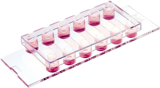
µ-Slide VI 0.4
- 可平行測試 6 種實驗條件,每個 channel 僅需 0.9–2.1 x 10⁴ 細胞與 30 µl 試劑,有效節省珍貴藥劑及抗體用量。
- 從細胞培養、染色到顯微鏡觀察,一氣呵成使用超便利!
- 微通道結構可避免因表面張力產生弧形液面的現象,細胞影像清晰不變形。
- 可彈性搭配針筒式、蠕動式、氣動式幫浦使用,模擬血管壁環境,應用於流體環境細胞培養 (搭配 ibidi Pump 使用,可大幅減少細胞與培養液用量)。
- 適合活細胞影像觀察攝影 (live cell imaging)、免疫螢光染色 (immunofluorescence)、流體環境細胞培養 (cell culture under flow) …等實驗使用。
歡迎與我們聯繫索取更多 µ-Slide VI 0.4 產品資訊與文獻。
產品特點
從細胞培養、染色到顯微鏡觀察“All-in-one”完成,一氣呵成高效便利

The ibidi Channel Slides (e.g., µ-Slide VI 0.1 | µ-Slide VI 0.4 | µ-Slide VI 0.5 Glass Bottom | µ-Slide I Luer) are particularly suitable for immunofluorescence stainings. The geometry of the ibidi Channel Slides is ideal for the exact exchange of small medium amounts, which is necessary during immunocytochemistry stainings. In addition, the coverslip bottom of the channel μ-Slides eliminates the need for additional coverslips.
微通道結構可避免因表面張力產生弧形液面的現象,光線可筆直穿透,整個觀察視野亮度均勻,影像清晰不變形
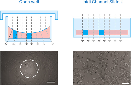
Open well: The meniscus is disturbing the phase contrast effect. Only the center provides convenient contrast. ibidi Channel Slides: In a channel geometry the beam path is always aligned. Phase contrast microscopy is possible independent from the location.
底部厚度符合 #1.5 蓋玻片,影像清晰易觀測
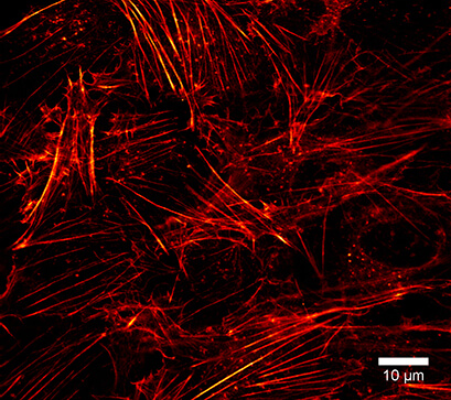
Super-Resolution Microscopy (STED) of the Actin Cytoskeleton. The µ-Slide VI 0.4 is compatible with super-resolution microscopy methods, such as simulated emission depletion (STED) microscopy. Using LifeAct-TagGFP2 Protein, the actin cytoskeleton can be visualized in detail. In this experiment, fixed Rat1 fibroblasts were incubated with LifeAct-TagGFP2 protein in a µ-Slide VI 0.4, ibiTreat. Microscopy was performed on the STEDYCON super-resolution STED nanoscopy system (Abberior Instruments GmbH, Göttingen, Germany) with a Plan-Neofluar 100x/1.4 objective lens.
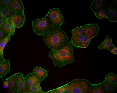
Fluorescence Microscopy for Mitochondria Visualization. The µ-Slide VI 0.4 is compatible with a variety of fluorescence microscopy methods. In this experiment, Madin-Darby Canine Kidney (MDCK) cells were cultured in a µ-Slide VI 0.4 and their mitochondria were visualized using MitoTracker. Image key: Mitochondria (MitoTracker, red), Actin cytoskeleton (Phalloidin, green), nuclei (DAPI, blue).
產品規格
µ-Slide VI 系列產品規格比較
 µ-Slide VI flat |
 µ-Slide VI 0.1 |
 µ-Slide VI 0.4 |
 µ-Slide VI 0.4 Bioinert |
 µ-Slide VI 0.5 Glass Bottom |
|
|---|---|---|---|---|---|
| Immunofluorescence assays | ✓ | ✓ | ✓ | ✓ | ✓ |
| Flow assays (cell culture under flow) | ✕ | ✓ | ✓ | ✓ | ✓ |
| 3D cell culture | ✕ | ✕ | ✕ | ✓ | ✕ |
| Outer dimensions | W25.5 x L75.5 mm | ||||
| Number of channels | 6 | ||||
| Channel length | 17 mm | ||||
| Channel width | 3.8 mm | 1 mm | 3.8 mm | 3.8 mm | 3.8 mm |
| Channel height | 0.4 mm | 0.1 mm | 0.4 mm | 0.4 mm | 0.54 mm |
| Channel volume | 30 µl | 1.7 µl | 30 µl | 30 µl | 40 µl |
| Growth area per channel | 0.6 cm² | 0.17 cm² | 0.6 cm² | 0.6 cm² | 0.6 cm² |
| Coating area per channel | 1.2 cm² (using 30 µl) |
0.37 cm² (using 1.7 µl) |
1.2 cm² (using 30 µl) |
1.2 cm² (using 30 µl) |
1.2 cm² (using 40 µl) |
| Volume per reservoir | None | 60 µl | 60 µl | 60 µl | 60 µl |
| Adapters | None | Female Luer | Female Luer | Female Luer | Female Luer |
| Bottom | ibidi Polymer Coverslip |
ibidi Polymer Coverslip |
ibidi Polymer Coverslip |
ibidi Polymer Coverslip with Bioinert surface |
Glass Coverslip No. 1.5H |
底部材質規格比較
| #1.5 ibidi Polymer Coverslip (ibidi 特殊塑料) |
#1.5H ibidi Glass Coverslip (#1.5H 高精度玻璃) |
|
|---|---|---|
| Bottom thickness | 180 µm (+10/-5 µm) | 170 µm (+/-5 µm) |
| Bottom material | Polymer | D 263 M Schott high precision glass |
| Gas permeability | Yes | No |
| Compatibility with protein coatings | Yes | Yes |
| Immersion oil compatibility | Yes | Yes |
| Autofluorescence | Low | Low |
| Transmission | Very high (even ultraviolet) | High (ultraviolet restrictions) |
| Refractive index (nD 589 nm) | 1.52 | 1.52 |
| Brightfield Microscopy | ++ | ++ |
| Phase Contrast | ++ | ++ |
| Differential Interference Contrast (DIC) | ++ | ++ |
| Widefield Fluorescence | ++ | ++ |
| Confocal Fluorescence | ++ | ++ |
| Total Internal Reflection Fluorescence (TIRF) | + | ++ |
| Super-Resolution Microscopy | + | ++ |
※更多材質特性比較資訊請見這裡。
訂購資訊
| Product Name | Surface | Pack Size | Cat. No. |
|---|---|---|---|
| µ-Slide VI 0.4 | ibiTreat: #1.5 polymer coverslip, tissue culture treated, sterilized | 15 | IB-80606 |
| 90 | IB-80606-90 | ||
| Collagen I: #1.5 polymer coverslip, sterilized | 15 | IB-80609 | |
| Collagen IV: #1.5 polymer coverslip, sterilized | 15 | IB-80602 | |
| Poly-L-Lysine: #1.5 polymer coverslip, sterilized | 15 | IB-80604 | |
| Uncoated: #1.5 polymer coverslip, hydrophobic, sterilized | 15 | IB-80601 |


