µ-Slides With Multi-Cell µ-Pattern
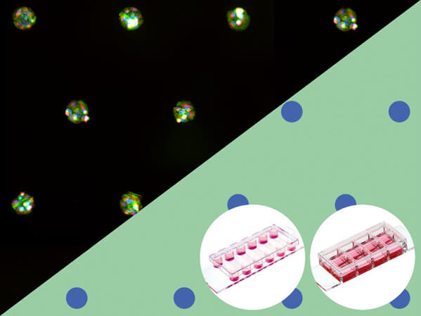
- 可使球狀體細胞 (spheroids)、類器官 (organoids) 間隔排列生長於玻片上,進行 3D 細胞培養與影像觀察。
- 具有 µ-Slide VI 0.4 與 µ-Slide 8 Well high 兩種產品款式可選 。
- µ-Slide VI 0.4 搭配 ibidi Pump 使用,可進行流體環境 3D 細胞培養 (3D cell culture under flow)。
- 由低自發螢光材質製成,且底部厚度符合 #1.5 蓋玻片標準,可直接結合高倍率光學或螢光顯微鏡進行細胞影像觀察。
本產品亦可提供客製化服務,歡迎與我們聯繫索取詳細資訊。
技術原理
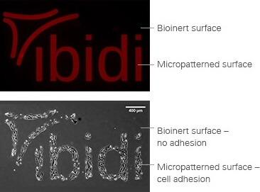
ibidi µ-Patterning 技術原理是先將一般 µ-Slide VI 0.4 或 µ-Slide 8 Well high 產品底部以特殊材質「Polyol hydrogel」處理,使其形成完成不具附著性的 Bioinert 表面。接著以共價鍵結的方式將源自於纖連蛋白 (Fibronectin) 結合區域的 RGD 三胜肽 (Arg-Gly-Asp, RGD tripeptide) 固定在 Bioinert 表面,使其形成供細胞附著生長的區域;根據 RGD 鍵結區域的大小與排列,可形成各種不同圖案設計。以上圖為例,紅色 ibidi 字樣為表面鍵結有 RGD peptides 的 µ-Pattern 區域,黑色則為不具有附著性的 Bioinert 區域,可以觀察到細胞僅會在 µ-Pattern 區域附著生長。
產品特點
µ-Pattern 特寫
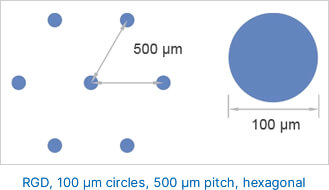
100 µm 直徑大小的圓形 RGD 鍵結區域以六角形網格排列於 Bionert 表面 (彼此間距 500 μm)。
底部厚度符合 #1.5 蓋玻片,影像清晰易觀測
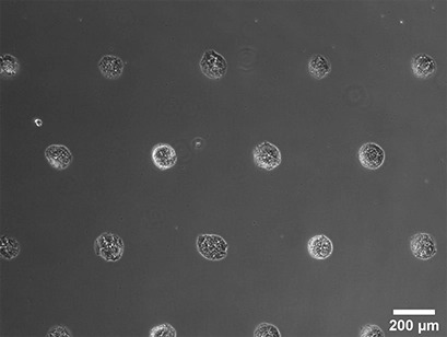
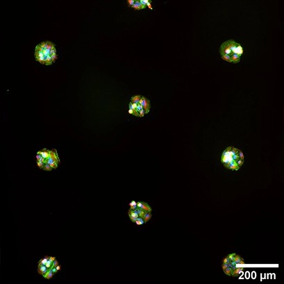
Spheroid formation of NIH-3T3 cells (murine embryo fibroblasts), 14 days after seeding single cells in the µ-Slide VI 0.4 With Multi-Cell µ-Pattern. Phase contrast microscopy, 4x objective lens.
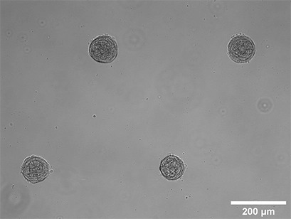
Spheroid formation of NIH-3T3 cells (murine embryo fibroblasts) 14 days after seeding single cells in the µ-Slide 8 Well With Multi-Cell µ-Pattern. Brightfield microscopy, 10x objective lens.
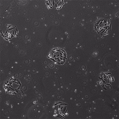
CAR-T Cell Killing Assays. RCC-26 cancer cells immobilized on multi cell pads. Effector T cells applied in a collagen I matrix (Collagen Type I, Rat Tail) induce apoptotic body formation of cancer cells.
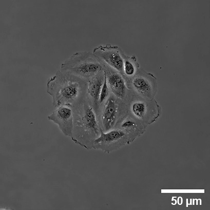
Live cell imaging of RCC-26 (human renal carcinoma) cells for 24 hours in the µ-Slide VI 0.4 With Multi-Cell µ-Pattern using the ibidi Stage Top Incubator. Phase contrast microscopy, 20x objective lens.
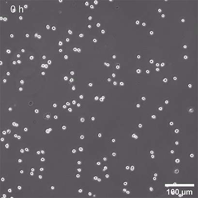
RCC-26 (human renal carcinoma) cells 24 hours after seeding in the µ-Slide VI 0.4 With Multi-Cell µ-Pattern, low density seeding for 2D monolayer. Phase contrast microscopy, 60x objective lens.
Immunofluorescence staining of RCC-26 (human renal carcinoma) cells 24 hours after seeding in the µ-Slide VI 0.4 With Single-Cell µ-Pattern. Green: F-actin (phalloidin), red: α-Tubulin, blue: nuclei (DAPI). Widefield fluorescence microscopy, 60x objective lens.
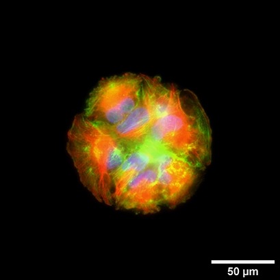
Immunofluorescence staining of RCC-26 (human renal carcinoma) cells 24 hours after seeding in the µ-Slide VI 0.4 With Multi-Cell µ-Pattern. Green: F-actin (phalloidin), red: α-Tubulin, blue: nuclei (DAPI). Widefield fluorescence microscopy, 10x objective lens.
產品規格
| µ-Slide geometry | See product page µ-Slide VI 0.4 or µ-Slide 8 Well high |
| Binding motif | RGD |
| Pattern shape | Circle |
| Diameter | 100 µm |
| Pitch | 500 μm |
| Pattern layout | Hexagonal |
| Number of patterns in µ-Slide VI 0.4 | ca. 300 per channel |
| Number of patterns in µ-Slide 8 Well high | ca. 450 per well |
| Surface passivation | Bioinert |
訂購資訊
| Product Name | µ-Pattern | Surface | Pack Size | Cat. No. |
|---|---|---|---|---|
| μ-Slide VI 0.4 μ-Pattern RGD, cir100, pit500, hex | RGD, 100 µm circles, 500 µm pitch, hexagonal | #1.5 polymer coverslip, micropatterned surface, surface passivation with Bioinert, sterilized | 2 | IB-83602-S |
| 10 | IB-83602 | |||
| μ-Slide 8 Well high μ-Pattern RGD, cir100, pit500, hex | RGD, 100 µm circles, 500 µm pitch, hexagonal | #1.5 polymer coverslip, micropatterned surface, surface passivation with Bioinert, sterilized | 2 | IB-83802-S |
| 10 | IB-83802 |
哪些細胞適用 µ-Pattern RGD 產品?
基本上只要能在 Fibronectin 處理表面生長的細胞,都能適用於 µ-Pattern RGD 產品,目前測試過的細胞包含:A549 (human lung carcinoma), ARPE-19 (human retinal pigmented epithelium), BEAS-2B (human lung epithelium), CHO (Chinese hamster ovary), HEK-293 (human embryonic kidney), HeLa (human cervical carcinoma), HT-1080 (human fibrosarcoma), HuH-7 (human liver carcinoma), L929 (murine fibroblasts), MCF-10A (human breast epithelium), MDA-MB-231 (human breast carcinoma), MDA-MB-436 (human breast carcinoma), MDCK.2 (canine kidney cells), NCI-H441 (human lung adenocarcinoma), NIH-3T3 (murine fibroblasts), RCC-26 (human renal carcinoma)。建議可先購買 2 片小包裝產品,測試您細胞在 µ-Pattern RGD 產品的生長情形。
更多款式選擇
 |
µ-Slides With Single-Cell µ-Pattern可使細胞以單細胞型式間隔排列生長於玻片上,便於評估藥物或 CAR-T 細胞活性 |
 |
µ-Slides With Test µ-Patterns單一玻片含多種不同表面間隔圖案設計,以便測試最佳適用類型 |


