µ-Slide Spheroid Perfusion
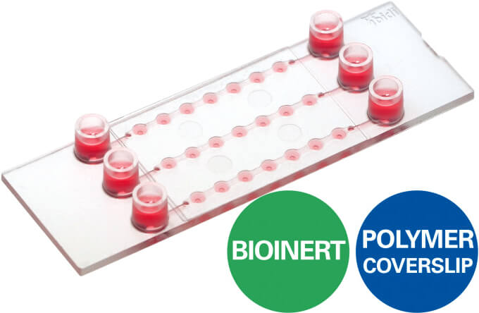
- 三條獨立微通道,每條通道含 7 個培養槽,搭配 ibidi Pump 使用即可進行流體環境 3D 細胞培養實驗 (3D cell culture under flow)。
- Bioinert 底部經特殊材質「Polyol hydrogel」處理,比一般 ULA (ultra low sttachment) 培養盤更加難以令細胞附著,特別適合球狀體細胞 (spheroids)、類器官 (organoids) 的長時間培養與觀測。
- 上蓋與底部厚度皆符合 #1.5 蓋玻片標準,可直接結合高倍率光學或螢光顯微鏡進行細胞影像觀察。
歡迎與我們聯繫索取更多 µ-Slide Spheroid Perfusion 產品資訊與文獻。
產品特點
µ-Slide Spheroid Perfusion 可搭配 ibidi Pump 使用,於流體環境進行 3D 細胞培養實驗
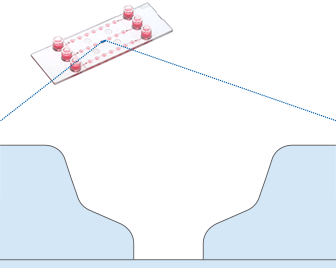
Optimal Nutrition Through Specialized Geometry. The µ-Slide Spheroid Perfusion is a specialized flow chamber for culturing free-floating 3D aggregates. It consists of 3 x 7 flat-bottomed wells, which are connected through a channel above. Each well forms its own niche, where the specimen is cultured. When applying perfusion through the channel, fresh medium continuously diffuses to the specimen. This ensures optimal nutrition and oxygen diffusion throughout the experiment without that the specimen is exposed to significant shear forces. The setup guarantees maximum of viability, but with minimum of shear stress for spheroids, organoids, or tissue.
µ-Slide Spheroid Perfusion 應用彈性多元
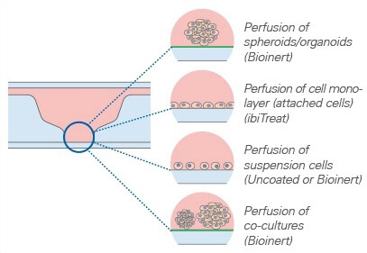
µ-Slide Spheroid Perfusion is available with three different surfaces (Bioinert, ibiTreat, Uncoated), and is suitable for a variety of applications.
µ-Slide Spheroid Perfusion 上蓋與底部厚度皆符合 #1.5 蓋玻片,影像清晰易觀測
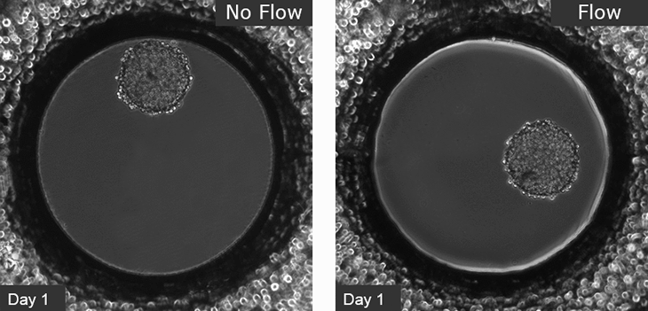
Highly Improved Spheroid Growth Rates When Cultured Under Perfusion. L929 fibroblasts show spheroid formation in the µ-Slide Spheroid Perfusion, Bioinert, days 1–14, seeding concentration 5 x 10⁵ single cells/ml. Left: no perfusion, medium exchange every second day. Right: perfusion with the ibidi Pump System, 0.75 ml/min. Phase contrast microscopy, 10x objective lens, well diameter 800 µm.
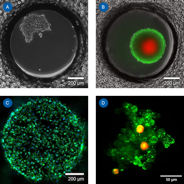
(A) Spheroid Formation of MCF-7 Breast Cancer Cells. Single cell seeding of MCF-7 breast cancer cells with spheroid formation in the µ-Slide Spheroid Perfusion, Bioinert, 1 day after seeding. Seeding concentration 5 x 10⁵ cells/ml. Phase contrast microscopy, 10x objective lens. (B) Live/Dead Staining of a Long-Term Cultured Spheroid. Live/dead FDA/PI staining of an L929 spheroid in the µ-Slide Spheroid Perfusion, Bioinert, after 14 days in culture with perfusion using the ibidi Pump System, 0.75 ml/min. Green: living cells (fluorescein diacetate, FDA); red: dead cells (propidium iodide, PI). Widefield fluorescence microscopy, 10x objective lens. (C) Fluorescence Microscopy of Adherent Cells. Fluorescence imaging of L929 fibroblasts in the µ-Slide Spheroid Perfusion, ibiTreat, fixated 2 hours after seeding. Green: F-actin (phalloidin); blue: nuclei (DAPI). Widefield fluorescence microscopy, 10x objective lens. (D) Organoid Co-Culture of PDAC Cells and Fibroblasts*. Organoid co-culture of the human pancreatic cancer (PDAC) cell line PA-TU-8988T (green, stained with CellTracker™ Green) and the murine fibroblast cell line mPSC4 (red, stained with CellTracker™ Orange CMTMR) in the µ-Slide Spheroid Perfusion. The µ-Slide was covered with a 25 µm FEP foil for matching the refractive index closer to water during upright light sheet microscopy. *The image was acquired by S. Volkery at MPI Muenster with the M Squared Aurora Airy beam upright light sheet setup. The sample was provided by K. Roth, University Marburg, Germany.
產品規格
| Outer dimensions | W25.5 x L75.5 mm |
| Adapters | Female Luer |
| Number of channels | 3 |
| Number of wells | 3 x 7 |
| Volume per well | 3.5 μl |
| Well height (bottom niche) | 0.4 mm |
| Well height (total) | 1.3 mm |
| Well diameter (bottom) | 0.8 mm |
| Channel volume (total) | 45 μl |
| Channel height | 0.2 mm |
| Channel width | 1.0 mm |
| Growth area per well | 0.5 mm² |
| Coating area using 3.5 µl | 9.7 mm² |
| Volume per reservoir | 60 μl |
| Top cover | ibidi Polymer Coverslip |
| Bottom | ibidi Polymer Coverslip |
訂購資訊
| Product Name | Surface | Pack Size | Cat. No. |
|---|---|---|---|
| µ-Slide Spheroid Perfusion | Bioinert: #1.5 polymer coverslip, surface passivation with Bioinert, sterilized | 15 | IB-80350 |
| ibiTreat: #1.5 polymer coverslip, tissue culture treated, sterilized | 15 | IB-80356 | |
| Uncoated: #1.5 polymer coverslip, hydrophobic, sterilized | 15 | IB-80351 |
流體環境 3D 細胞培養產品比較表
 µ-Slide Spheroid Perfusion |
 µ-Slide III 3D Perfusion |
 µ-Slide I Luer 3D |
|
|---|---|---|---|
| Perfusion of samples | ✓ | ✓ | ✓ |
| Defined shear stress on cell monolayers | ✕ | ✕ | ✓ (on gel matrix) |
| Gel matrices for 3D | ✕ | ✓ | ✓ |
| Spheroids/organoids | free floating in well | inside gel matrix only | inside gel matrix only |
| Suspension cells | free floating in well | inside gel matrix only | inside gel matrix only |
| Adherent cells | on coverslip | inside or on gel matrix | inside or on gel matrix |


