CytoSMART Lux3 BR 活細胞即時影像監測系統
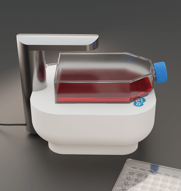 體型輕巧便利的 CytoSMART Lux3 BR 能讓您隨時隨地掌握細胞生長狀態與進行數據分析!其優勢特點包含:
- 機身小,可直接放入培養箱,即時觀察細胞型態。
- 可拍攝高達 2027x2027 pixels 清晰照片,輕鬆取得高解析度影像。
- 內建影像分析,可進行細胞滿度、爬行、Spheroid/Colony cell 偵測等分析。
- 可設定 5 分鐘~12 小時拍攝間隔,並可持續拍攝長達數週影像,支援縮時影片製作。
- 影像即時上傳雲端,不用進實驗室,在家就能即時監控細胞生長情況。
- 最多可四台連線,一次控制,擴大分析資料量。
- 不論是 Plates、Dishes、Flasks 或微流體晶片皆可適用。
- 支援 Open API,可以輕易和自動控制系統整合。
歡迎與我們聯繫索取更多 CytoSMART Lux3 BR 產品資訊與文獻。
產品特點
可拍攝高達 2027x2027 pixels 清晰照片,輕鬆取得適用於文獻發表與實體印刷的高解析度影像
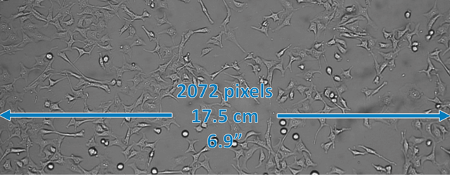
Fig. 1 | Digital images suitable for scientific publications. The CytoSMART Lux3 BR is ideal for capturing crisp brightfield images and videos of living cells. The image size of 2072×2072 pixels combined with the 1.45×1.45 mm field of view provide a resolution of 0.7 µm/pixel. Even at the commonly required image resolution of 300 dpi for printed scientific publications, these images can fill the entire page width if desired, without compromising the image quality.
CytoSMART Lux3 BR 應用於細胞趨化性實驗 (Chemotaxis assay) 的實際案例數據
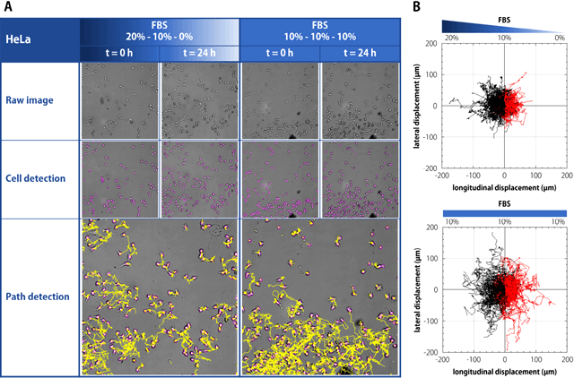
Fig. 2 | HeLa cells were seeded in the ibidi µ-Slide Chemotaxis, with either FBS gradient or constant FBS concentration. Cultures were monitored for 24 h using the CytoSMART Lux3 BR Duo Kit, with an imaging interval of 5 min. HeLa cells cover a larger distance in constant FBS concentration, but prefer to migrate towards a higher FBS concentration when exposed to a gradient. (A) Raw images of HeLa cells made using CytoSMART Lux3 BR Duo Kit, single cell detection (purple) and path detection (yellow) with FIJI-plugin TrackMate. (B) Chemotactic displacements of HeLa cells visualized using FIJI-plugin Chemotaxis and Migration Tool, with longitudinal and lateral displacements defined with respect to the FBS gradient.
產品規格
| Channel | Brightfield, with digital phase contrast |
| Camera | 6.4 MP CMOS |
| Magnification | 10× fixed objective, additional 2× digital zoom |
| Image size | 2072 x 2072 pixels |
| Field of view | 1.45 x 1.45 mm |
| Light source | LED |
| Data formats | JPEG, TIFF, XLSX, MP4 |
| Data storage | Unlimited cloud storage |
| Computer requirements | Windows 10, USB 3.0, internet, 8 GB RAM, 256 GB SSD |
| Dimensions | L166 x W140 x H135 mm |
| Weight | 1.3 kg |
| Culture vessels | Well-plates, petri dishes, flasks, microfluidic chips and custom culture vessels |
| Operating conditions | 5–40 °C, 20–95% humidity |
| Warranty | 1 year parts and labor |
| Extra features | Confluence algorithm, scratch assay analysis algorithm |
訂購資訊
| Product Name | Cat. No. |
|---|---|
| CytoSMART Lux3 BR | O-KAB-1009 |


