|
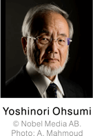 以 2016 年諾貝爾生理學或醫學獎得主大隅良典 (Yoshinori Ohsumi) 的研究發現為基礎,科學家們終於得以深入探討細胞自噬 (Autophagy) 與人類疾病之間的關係。目前已知,細胞自噬與第二型糖尿病、神經退化性疾病、癌症等多種疾病皆具有相關性,透過尋找具有調控細胞自噬功能的藥物,極有可能找出能夠緩解或治療疾病的有效途徑。 以 2016 年諾貝爾生理學或醫學獎得主大隅良典 (Yoshinori Ohsumi) 的研究發現為基礎,科學家們終於得以深入探討細胞自噬 (Autophagy) 與人類疾病之間的關係。目前已知,細胞自噬與第二型糖尿病、神經退化性疾病、癌症等多種疾病皆具有相關性,透過尋找具有調控細胞自噬功能的藥物,極有可能找出能夠緩解或治療疾病的有效途徑。
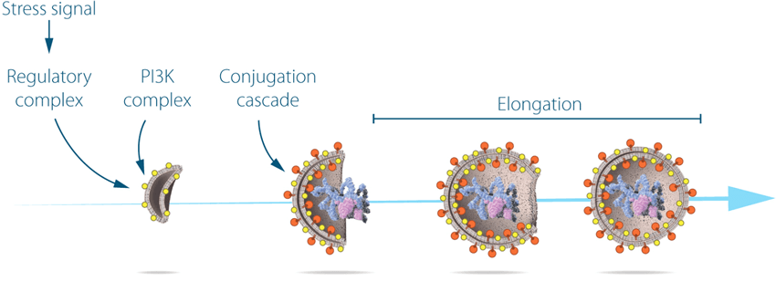
Ohsumi studied the function of the proteins encoded by key autophagy genes. He delineated how stress signals initiate autophagy and the mechanism by which proteins and protein complexes promote distinct stages of autophagosome formation. IMAGE © Nobel Media AB 2018.
細胞自噬高效檢測工具 — 以 p62 偵測為實例
目前細胞自噬活性偵測多以顯微影像觀察為主,例如透過觀察標幟有螢光分子的細胞自噬作用指標蛋白 LC3 或 p62,於藥物干預前後在細胞內的蛋白質表現量增減、以及分佈情形變化,來瞭解藥物對細胞自噬作用的影響性。此種偵測方式,在面對藥物庫篩選或大量樣本分析時,就顯得較為耗時而費力。
國際知名新藥研發技術開發商 PerkinElmer 在其最新發表的技術專文中,使用 AlphaLISA 技術對 p62 進行定量偵測。AlphaLISA 技術原理類似 ELISA,但卻擁有優於 ELISA 的偵測靈敏度、以及免洗滌﹑步驟簡便的雙重優勢。使用 AlphaLISA 技術,不僅節省樣本用量 (5 μL/well),並且可於 2 小時快速完成大量樣本的分析試驗。
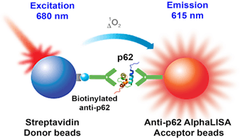 AlphaLISA p62 assay principle. A biotinylated anti-p62 antibody is coupled to a Streptavidin Donor bead, while an anti-p62 antibody is coupled to an Acceptor bead. Binding p62 brings the Acceptor and Donor bead in close proximity, so that when the Donor bead is excited at 680 nm, the Acceptor bead produces an emission at 615 nm. The AlphaLISA p62 detection assay generates results in under two hours, with no-wash steps. Image from Detection and Quantification of Autophagy Using the AlphaLISA p62 Assay. PerkinElmer. Fig 1. AlphaLISA p62 assay principle. A biotinylated anti-p62 antibody is coupled to a Streptavidin Donor bead, while an anti-p62 antibody is coupled to an Acceptor bead. Binding p62 brings the Acceptor and Donor bead in close proximity, so that when the Donor bead is excited at 680 nm, the Acceptor bead produces an emission at 615 nm. The AlphaLISA p62 detection assay generates results in under two hours, with no-wash steps. Image from Detection and Quantification of Autophagy Using the AlphaLISA p62 Assay. PerkinElmer. Fig 1. |
 AlphaLISA p62 Assay AlphaLISA p62 Assay
文章中使用 HeLa 細胞作為研究對象,將細胞分別培養在不含或含有自噬作用抑制劑 Chloroquine (50 μM) 的培養液內 18 小時。移除培養液後,先以 PBS 清洗細胞兩次,接著使用 50 μL AlphaLISA 細胞裂解液溶解細胞。
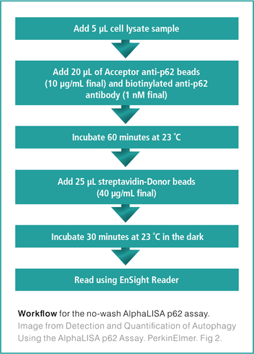
將細胞溶解物 (cell lysates) 加入 AlphaPlate™-384 微量多孔盤中 (5 μL/well),與 anti-p62 AlphaLISA Acceptor beads、biotintylated anti-p62 antibody 於室溫下作用反應 1 小時,然後加入 Streptavidin Donor beads 避光作用半小時,即可進行訊號讀取。詳細流程步驟說明,請見右方圖示。
AlphaLISA p62 Assay 偵測結果顯示,Chloroquine 會引致細胞內 p62 表現量的增加。為驗證 AlphaLISA 實驗結果,作者以細胞免疫螢光染色實驗證實,加入 Chloroquine 處理後的細胞的確可觀察到 p62 蛋白表現量明顯提高的現象。文中,作者進一步使用 Hoechst 染色活細胞計數結果來校正 AlphaLISA 檢測數值,使實驗結果更精準可信。
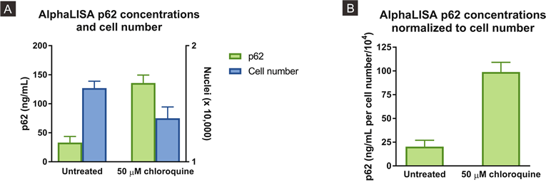
(A) AlphaLISA p62 concentrations and cell number for each condition and (B) AlphaLISA p62 concentrations normalized to cell number. Image from Detection and Quantification of Autophagy Using the AlphaLISA p62 Assay. PerkinElmer. Fig 6. |
| |
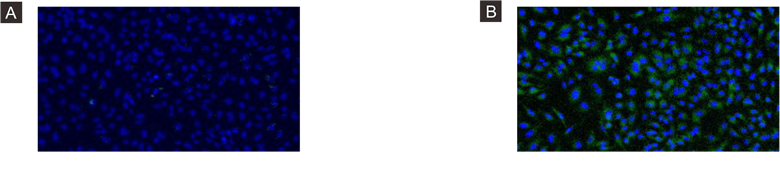
Representative EnSight images of (A) untreated HeLa cells or (B) with treatment of 50 µM chloroquine. Hoechst staining is in blue and FITC labeled p62 is in green. Image from Detection and Quantification of Autophagy Using the AlphaLISA p62 Assay. PerkinElmer. Fig 5. |
透過本文,我們可以發現 AlphaLISA 能夠取代傳統 ELISA,以更精簡的步驟﹑更高的偵測靈敏度完成大量樣本檢測。相較於傳統細胞螢光影像的自噬活性檢測方式,AlphaLISA 不會受制於繁瑣的染色步驟、以及免疫螢光實驗流程中常見的長時間培養等問題,能夠顯著縮短獲取實驗結果的時間,並且適用於各種細胞類型,可作為細胞自噬研究的高效檢測工具。
參考資料
- The Nobel Prize in Physiology or Medicine 2016. NobelPrize.org. Nobel Media AB 2018. Thu. 18 Oct 2018. [Summary] [Press release]
- 李岳倫 (2016) 諾貝爾生醫獎大隅良典與細胞自噬. 科學月刊. [Article]
- Bhutia SK (2018) Monitoring and Measuring Mammalian Autophagy. Methods Mol Biol. 1854:209-222. PubMed: 29855817
- Yoshii SR (2017) Monitoring and Measuring Autophagy. Int J Mol Sci. Aug 28;18(9). pii: E1865. PubMed: 28846632
- Detection and Quantification of Autophagy Using the AlphaLISA p62 Assay. PerkinElmer. [Application Note]
|